This repository contains example code from the paper in preparation on preclinical cancer imaging titled "MRI-based Deep Learning Segmentation and Radiomics of Sarcoma Tumors in Mice."
This work is part of the U24 co-clinical trial of which CIVM at Duke University is a participant. This work has been funded by NIH U24CA220245.
This goal of this project is to create an pipeline with two facets:
- Automatic segmentation of soft tissue sarcoma using deep learning.
- Automate calculation and analysis of radiomic features within the tumor population.
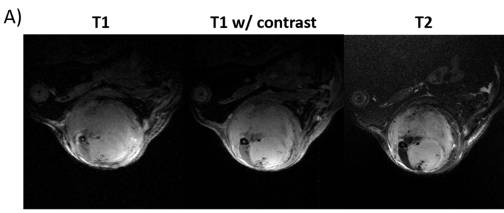
The data used in this study is composed of multi-contrast MR images of soft tissue sarcoma. Tumors were imaged using
T2-weighted and T1-weighted sequences. These were followed by a contrast-enhanced T1-weighted acquisition.

The full dataset will soon be available on the CIVM VoxPort page
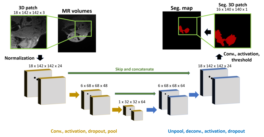
Segmentation was performed via a U-net CNN. The network functions on patches taken from image volumes. The general
network structure is shown below.

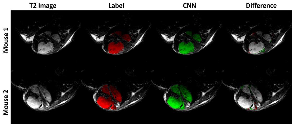
Training and perfomance anlysis is done using the Segmentation.py script. The results for a network trained on multi-contrast MR images with cross entropy loss is shown below.
T2-weighted images are bias corrected using N4BiasFieldCorrection in ANTs.
Network structures have been defined in model_keras.py. A variety of networks have been tested, including those with and without skip connections as well as testing out cross entropy and dice loss functions.
Radiomic features were calculated within the segmentation using PyRadiomics. Radiomic calculation are performed using radiomic_calculations_batch.py.
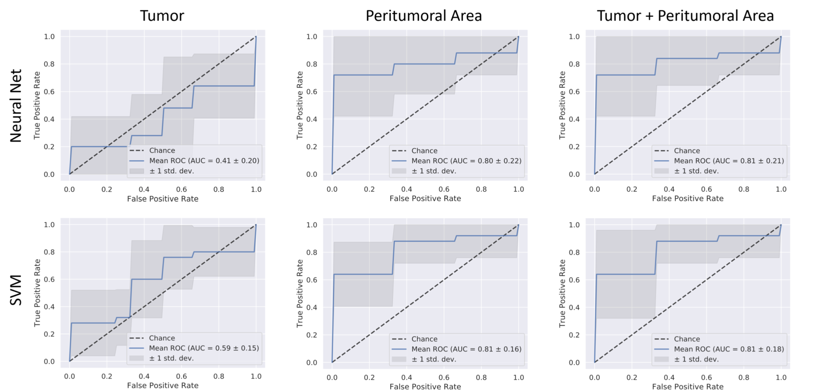
The code used to process the radiomic features is found in [radiomic_classifications.py] (Radiomics/radiomics_classifications.py). The features are plotted and classified using SVMs and NNs to determine if primary local recurrence can be predicted in this population based only on radiomic features. Machine learning classifier functions lie in radiomic_functions.py.
Using this code, we achieved an AUC of 0.81 for predicting recurrence within these mice.
Due to the high dimensionality of the data (321 features per tumor) feature selection is required . This are accomplished via mRMR, which must be added to the path before running.
A crawler has been implemented to locate, pre-process, segmentent, and compute radiomic features for large collections of images. The code for this located in the "Crawler" folder and can be run through the crawler.py script once the base paths have been updated.


