DICOM Imaging Router: An Open Deep Learning Framework for Classificationof Body Parts from DICOM X-ray Scans
This repository contains the training code for our paper entitled "DICOM Imaging Router: An Open Deep Learning Framework for Classificationof Body Parts from DICOM X-ray Scans", which was accepted by ICCV Workshop 2021 on Computer Vision for Automated Medical Diagnosis (CVAMD) .
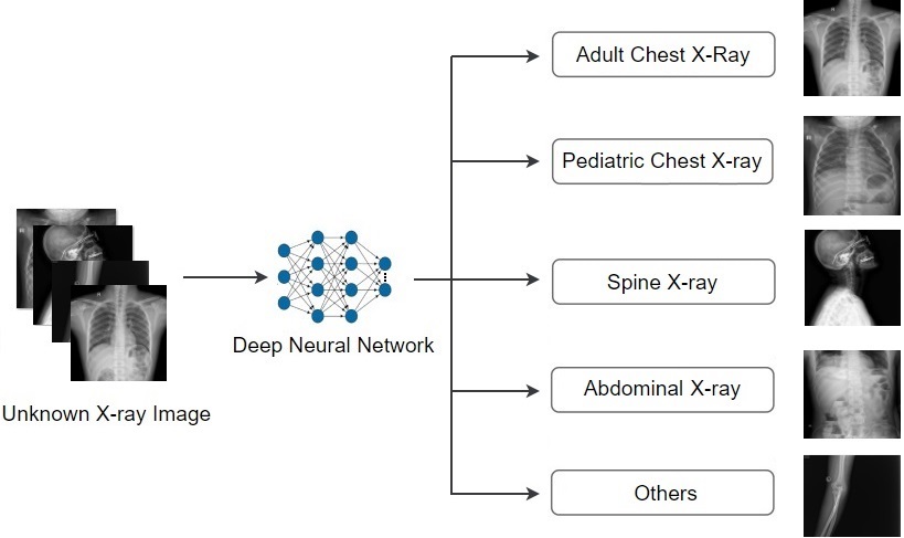
X-ray imaging in Digital Imaging and Communications in Medicine (DICOM) format is the most commonly used imaging modality in clinical practice, resulting in vast, non-normalized databases. This leads to an obstacle in deploying artificial intelligence (AI) solutions for analyzing medical images, which often requires identifying the right body part before feeding the image into a specified AI model. This challenge raises the need for an automated and efficient approach to classifying body parts from X-ray scans. Unfortunately, to the best of our knowledge, there is no open tool or framework for this task to date. To fill this lack, we introduce a DICOM Imaging Router that deploys deep convolutional neural networks (CNNs) for categorizing unknown DICOM X-ray images into five anatomical groups: abdominal, adult chest, pediatric chest, spine, and others. To this end, a large-scale X-ray dataset consisting of 16,093 images has been collected and manually classified. We then trained a set of state-of-the-art deep CNNs using a training set of 11,263 images. These networks were then evaluated on an independent test set of 2,419 images and showed superior performance in classifying the body parts. Specifically, our best performing model (i.e., MobileNet-V1) achieved a recall of 0.982 (95% CI, 0.977-0.988), a precision of 0.985 (95% CI, 0.975-0.989) and a F1-score of 0.981 (95% CI, 0.976-0.987), whilst requiring less computation for inference (0.0295 second per image). Our external validity on 1,000 X-ray images shows the robustness of the proposed approach across hospitals. These remarkable performances indicate that deep CNNs can accurately and effectively differentiate human body parts from X-ray scans, thereby providing potential benefits for a wide range of applications in clinical settings. The dataset, codes, and trained deep learning models from this study will be made publicly available on our project website at VinDr Datasets.
- Image resize (512,512)
- Split data: training, testing, valid dataset (0.7, 0.15, 0.15)
- The classes: abdominal, adult chest, pediatric chest, spine, and others.
- Configuaration used in the paper are in folder utils. It is recommended that you change training configuaration in constants.py
  step1:
    pip install -r requirements.txt
  step2:
    python training.py
  step3:
    python test.py
| Model | Recall | Precision | F1-score |
|---|---|---|---|
| MobileNet-V1 | 0.982(0.975-0.987) | 0.981(0.975-0.987) | 0.981(0.976-0.987) |
| MobileNet-V2 | 0.967(0.985-0.976) | 0.979(0.974-0.985) | 0.972(0.965-0.980) |
| ResNet18 | 0.923(0.909-0.937) | 0.939(0.927–0.951) | 0.930(0.917–0.942) |
| ResNet34 | 0.923(0.909–0.937) | 0.935(0.923–0.948) | 0.929(0.916–0.941) |
| EfficientNet-B0 | 0.975(0.968–0.981) | 0.980(0.975–0.986) | 0.977(0.971–0.983) |
| EfficientNet-B1 | 0.969(0.961–0.977) | 0.977(0.971–0.983) | 0.973(0.966–0.980) |
| EfficientNet-B2 | 0.973(0.965–0.980) | 0.977(0.972–0.984) | 0.975(0.969–0.982) |
